BACKGROUND

The purpose of the cardioversion is to interrupt the abnormal electrical circuit(s) in the heart and to restore a normal heartbeat. The delivered electrical shock causes all the heart cells to contract simultaneously, thereby interrupting and terminating the abnormal electrical rhythm (typically fibrillation of the atria) without damaging the heart. The heart’s electrical system then restores a normal heartbeat.

What is an Electrical Cardioversion?

Cardioversion is a corrective procedure where an electrical shock is delivered to the heart to convert, or change, an abnormal heart rhythm back to normal sinus rhythm. Most elective or “non-emergency” cardioversions are performed to treat atrial fibrillation (A Fib) or atrial flutter (AFL), non-life threatening abnormal rhythms in the top of the heart. Cardioversion is also used in emergency situations to correct an abnormal rhythm when it is accompanied by faintness, Low Blood Pressure, chest pain, difficulty breathing, or loss of consciousness. Sometimes a Transeophageal Echocardiogram is performed prior to an Electrical Cardioversion to exclude cardiac thrombus and clot.

Normal heart rhythm

In order for the heart to do its work (pumping blood throughout the body), it needs a sort of spark plug or electrical impulse to generate a heartbeat. Normally this electrical impulse begins in the upper right chamber of the heart (in the right atrium) in a place called the sino-atrial (SA) node. The SA node is the natural pacemaker of the heart. The SA node gives off electrical impulses to generate a heartbeat in the range of 60 to 100 times per minute. If you are exercising, doing strenuous work or you are under a lot of stress, your heart rate may be faster. When you rest or sleep your heart rate will slow down. If you take certain medications, your heart rate may be slower.

From the SA node, the electrical impulse is relayed along the heart’s conduction system. It spreads throughout both the right and left atria causing them to contract evenly. When the impulse spreads over the right atrium it reaches the atrio-ventricular (AV) node. This is a very important structure in the heart because it is the only electrical connection between the top chambers and the bottom chambers. It is therefore the only way in which an electrical impulse can reach the pumping chambers (the ventricles). The impulse spreads through the AV node and down into the lower chambers or ventricles of the heart. This causes them to contract and pump blood to the lungs and body.

What is Atrial Fibrillation?

Atrial fibrillation is due to the development of electrical short circuits inside the top chambers of the heart. Usually these short circuits begin in the top chamber on the left (left atrium). They are usually triggered by abnormal electrical activity located within the veins that drain blood from the lungs back to the heart (pulmonary veins)

These rapid short circuits have several consequences:

  1. The short circuits drive the pumping chambers rapidly and erratically. This produces palpitations, shortness of breath, and tiredness. In some people it can also cause dizzyness and chest pain. It usually reduces your exercise or physical capacity. It is NOT however a life-threatening rhythm disturbance.
  2. The short circuits result in ineffective pumping of the upper chambers. This leads to slow blood flow in both of these upper chambers (the left and right atria). This can rarely cause blood clots and possibly stroke. For this reason many (but not all) patients with atrial fibrillation will require a blood thinner. This may be aspirin, warfarin or one of the newer medications (Pradaxa, Xarelto or Eliquis).

What is Atrial Flutter?

Atrial Flutter is due to a short circuit in one of the upper chambers of your heart termed the right atrium. This rapid short circuit has several consequences:

  1. The short circuit drives the pumping chambers very rapidly and sometimes erratically. This produces palpitations, shortness of breath, and tiredness. In some people it can also cause dizziness and chest pain.
  2. The short circuit results in ineffective pumping of the upper chambers. This leads to slow blood flow in both of these upper chambers (the left and right atrium). This can rarely cause blood clots and possibly stroke. One of the major reasons to cure atrial flutter is to prevent this risk of stroke.

PROCEDURE INFORMATION

What happens during an Electrical Cardioversion?

The electrical cardioversion is performed in the angiography suite, coronary care unit or recovery room. It is necessary for you to fast for at least 6 hours prior to the cardioversion. You can take your usual medications on the morning of the procedure with a small amount of water. In many cases you will have been on warfarin prior to the cardioversion. It is important that the level of the blood thinning (the INR) is checked prior to the cardioversion, usually the day prior or the morning of the procedure.

Your cardioversion will only be performed if you have had a minimum of 3 weeks whereby your INR has been between 2 and 4. Some patients will be taking the new anticoagulant medications (called NOACs). These are not monitored with the INR so it is important that you tell us if you have not been taking blood-thinning medications regularly for the last 3 weeks.

If you have not been taking your blood thinning medications regularly it is vital that you notify the staff undertaking the cardioversion. Occasionally, a trans-esophageal echocardiogram (TOE) will be performed prior to cardioversion. This is usually done when there is a high index of suspicion of clots inside the heart or your warfarin level has been fluctuating or if you have not taken your NOAC medication regularly.

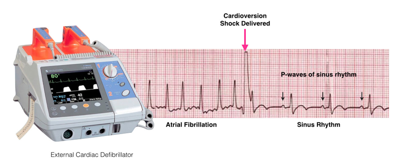
For the cardioversion, special pads are used to minimise any skin burning or irritation from the electrical shock. You will be connected to an external defibrillator to allow monitoring of your heart rhythm and to allow the application of the necessary energy to restore your heart’s rhythm back to normal. As the shock may be uncomfortable, some form of sedation or anesthesia is administered intravenously.

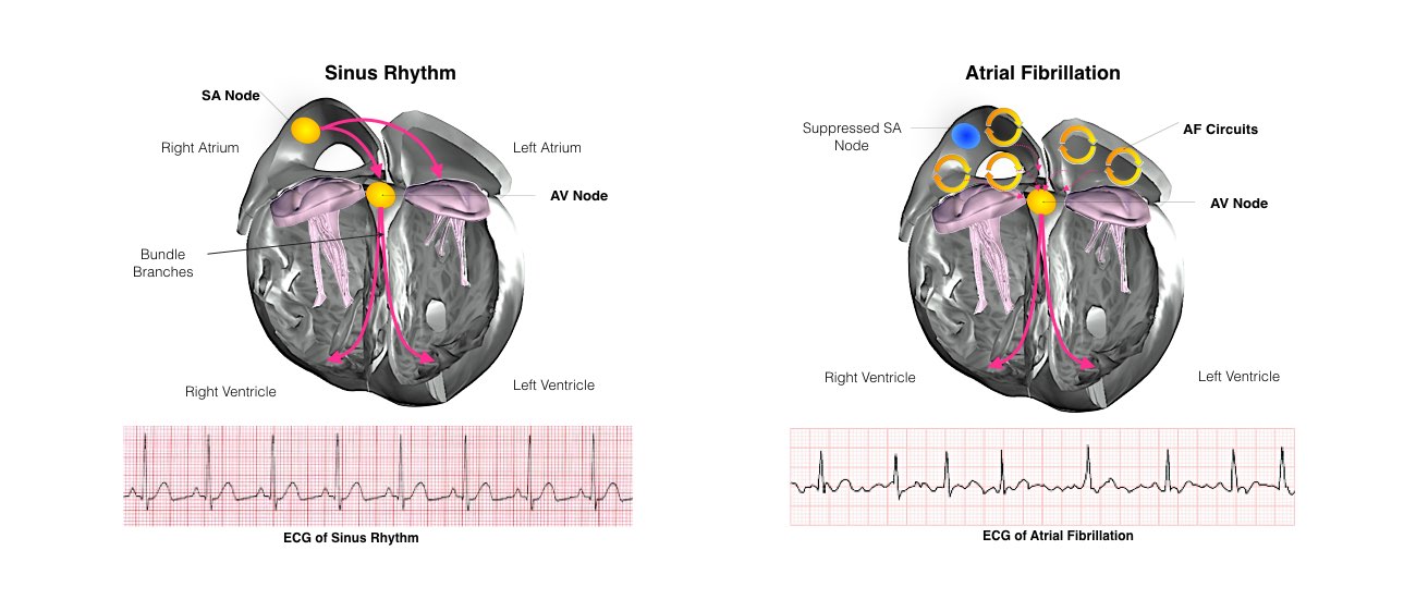
Once you are sedated, the shock is delivered. Additional shocks at higher energy levels can be delivered if the first shock does not restore the rhythm back to normal. The success rate depends upon a number of factors but there is an approximately 90% chance that the normal rhythm (sinus rhythm) will be restored.

There is a very small risk of stroke immediately after the cardioversion procedure as a result of stunning of the top chamber of the heart (atrium). This risk is greatest in the first few weeks after the cardioversion procedure. This is why it is crucial that warfarin is continued (maintaining the INR between 2 and 4) for at least a month after the procedure. For patients on NOACS, it is crucial you take your medications regularly for the next month.

In a proportion of patients, the arrhythmia subsequently recurs and repeated cardioversion may be required. Patients generally wake quickly and without any recollection of the shocks because of the effects of the sedatives.

You will not be able to drive yourself home from the procedure and will need a responsible adult to stay with you overnight after the procedure. You should not drive or make any important decisions for 24 hours after the procedure.

What is a transesophageal echocardiogram?

A transesophageal echocardiogram is a diagnostic test used to view the structures of the beating heart. The procedure is performed under conscious sedation prior to the electrical cardioversion procedure to make sure there are no blood clots in the heart.

In this procedure, a transducer is passed through the mouth and into the esophagus, which lies behind the heart. The transducer sends images of the heart to a monitor. This imaging process is called an echocardiogram. An echocardiogram uses ultrasound waves to create moving pictures of the structures of the heart such as the chambers and valves. In particular we want to make sure there are no blood clots in the heart prior to the cardioversion procedure.

PRE/POST PROCEDURE

How long will I be in hospital for after the Cardivoersion?

You will usually only need to go to hospital for part of the day, although some people need to stay overnight.

RISKS/BENEFITS

How successful are cardioversions?

Cardioversion doesn’t always restore normal heart rhythm. Sometimes it’s successful to start with, but then your abnormal heart rhythm comes back several days, weeks or even months later. If this happens, your doctor may want to repeat the cardioversion or they may consider another treatment for you.

Cardioversion Risks

Cardioversion is a very safe and effective treatment for atrial fibrillation and atrial flutter. However, there are some potential complications.

  • Stroke (or embolism) is perhaps the most dreaded complication. This is where a blood clot is dislodged from the heart into the blood stream. If the clot travels to a major organ it can cause problems such as a stroke or heart attack. For this reason if the atrial fibrillation has been present for more than 48 hours, or its duration is unknown, either the blood must be thinned using warfarin for at least 3 weeks or you must undergo TOE examination prior to the cardioversion.
  • If adequate blood thinning is undertaken the risk of an embolus is less than 1:1000.
  • Depending on how long you have been in atrial fibrillation or flutter, your normal pacemaker may not function immediately on terminating the abnormal rhythm. Usually this recovers promptly but in rare cases temporary and sometimes permanent pacemakers may need to be inserted.

Finally, there is a very small risk associated with the administration of the sedation required for the cardioversion, but the likelihood of a significant anaesthetic complication is also less than 1:1000.

Hi there!