BACKGROUND

The heart is a pump responsible for maintaining blood supply to the body. It has four chambers. The two upper chambers (the right atrium and left atrium) are the chambers that receive blood as it returns from the body via the veins. The lower chambers (the right and left ventricle) are the chambers responsible for pumping the blood out to the body via the arteries. Like any pump, the heart has an electrical system that controls how it functions.

Normal heart rhythm

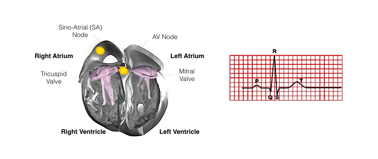
In order for the heart to do its work (pumping blood throughout the body), it needs a sort of spark plug or electrical impulse to generate a heartbeat. Normally this electrical impulse begins in the upper right chamber of the heart (in the right atrium) in a place called the sino-atrial (SA) node. The SA node is the natural pacemaker of the heart. The SA node gives off electrical impulses to generate a heartbeat in the range of 60 to 100 times per minute. If you are exercising, doing strenuous work or are under stress, your heart rate will be faster. When you rest or sleep your heart rate will slow down. If you take certain medications, your heart rate may be slower.

From the Sinus Node, the electrical impulse is relayed along the heart’s conduction system. It spreads throughout both the right and left atria causing them to contract evenly.
When the impulse spreads over the right atrium it reaches the atrio-ventricular (AV) node. This is a very important structure in the heart because it is the only electrical connection between the top chambers and the bottom chambers. It is therefore the only way in which an electrical impulse can reach the pumping chambers (the ventricles). The impulse spreads through the AV node and down into the lower chambers or ventricles of the heart. This causes them to contract and pump blood to the lungs and body.

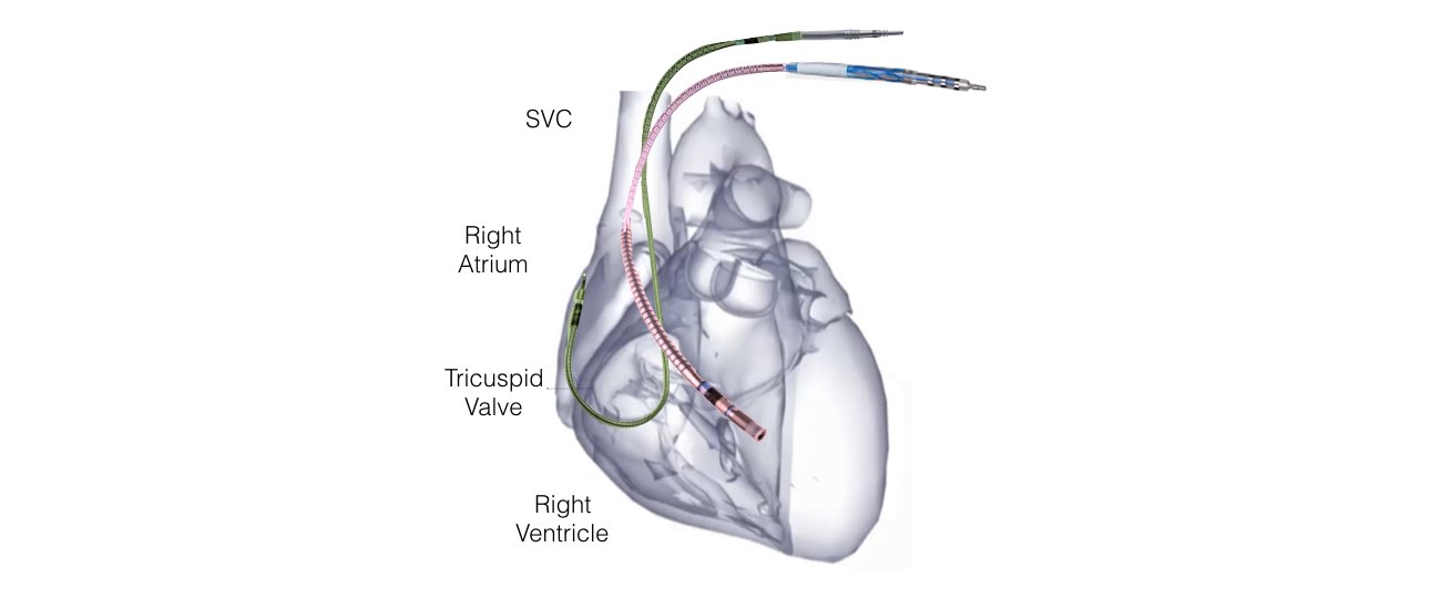
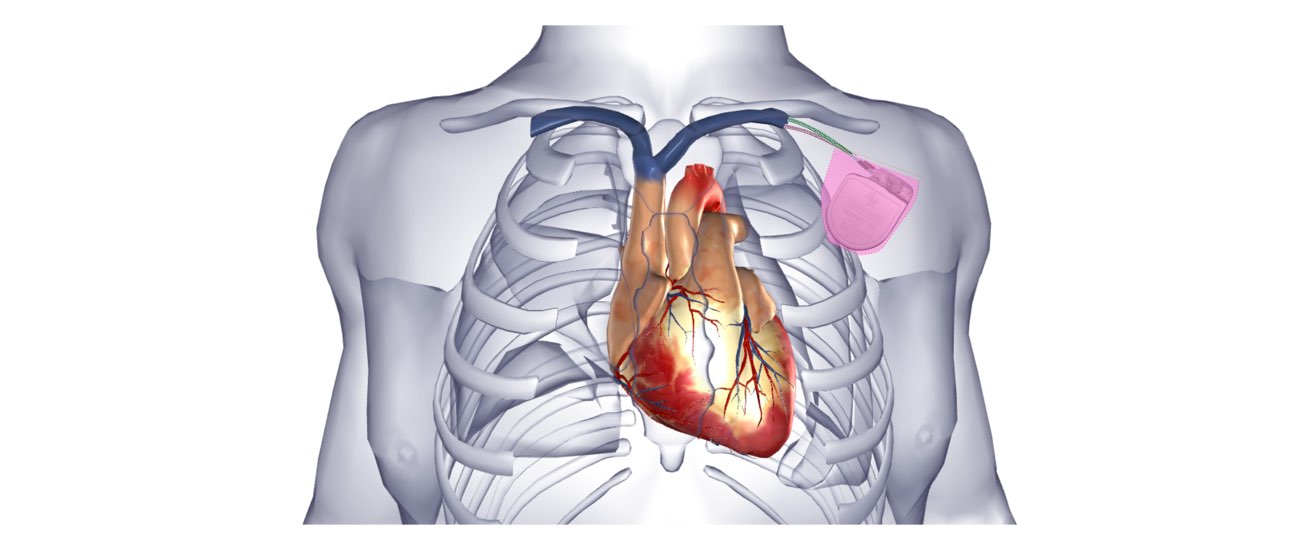
The main body of the device is usually implanted under the skin in your shoulder region. The leads are connected to the device at one end and to your heart at the other end. The leads are able to transfer information about your heart rhythm to the device. The device will determine what rhythm your heart is in and will deliver therapy through the leads when necessary.

Why do I need a defibrillator?

In some people the electrical system of the heart may develop a short circuit in the pumping chambers (ventricles). This short circuit is termed Ventricular tachycardia or ventricular fibrillation. These rhythm disturbances cause the heart to beat too rapidly and inefficiently so that not enough blood is pumped to the brain.

The usually symptoms are intense dizziness, or sudden collapse with loss of consciousness.

These heart rhythm disturbances are potentially fatal.

Your doctor is recommending you have a defibrillator either because you have had one of these rhythm disturbances or because you may be at increased risk of having this in the future.

If the life-threatening rhythm occurs, the defibrillator delivers a high energy shock (up to 750 volts) in order to return the heart to a normal rhythm. This is life-saving treatment.

What is an ICD?

An ICD, or Implantable Cardioverting Defibrillator, is a device implanted in the body that can detect and terminate fast heart rhythms (Ventricular Tachyarrhythmias). An ICD has two main parts”

  1. Main body (battery and electrical circuitry)
  2. Leads
learn more about heart conditions at one heart cardiology

PROCEDURE INFORMATION

What is involved in Defibrillator Implantation?

Insertion of a defibrillator is now a very common procedure. This is performed either under local anaesthetic with sedative medication to make you feel comfortable or under general anaesthetic. Your doctor will discuss this with you. The procedure takes approximately 1 hour and is performed in the cardiac catheter laboratory.

This is a special room that has a patient table, X-Ray tube, ECG monitors and other equipment. The staff in the lab will all be dressed in hospital theatre clothes and during the procedure will be wearing hats and masks.

Many ECG monitoring electrodes will be attached to your chest area. A nurse or doctor will insert an intravenous line usually into the back of your hand. This is needed as a reliable way to give you medications during the study without further injections. You will also have a blood-pressure cuff attached to your arm that will automatically inflate at various times throughout the procedure.

The defibrillator is inserted just under the collar-bone on the front of the chest usually on the left side. The area is prepared with a special sterile solution that may feel cold. You will be covered by a large sterile sheet that will partly cover your face. You will be able to look out from under the sheet to the side and a nurse will be present at all times. You will be given oxygen to breathe by a small tube that is positioned under your nostrils or by a mask that covers your face.

At the start of the procedure, the doctor will inject local anaesthetic into the area under the collar-bone where the defibrillator is to be inserted. This will sting momentarily but the area will then be numb. During the procedure you may feel some firm pushing in the shoulder area but this should not be painful. If you experience pain or discomfort you should tell the nurse or doctor.

Usually 2 leads (special wires which connect the defibrillator with the heart) are inserted into the heart via the vein under the collarbone. These are manipulated into the heart under X-Ray control. Most commonly 2 leads are inserted, one into the right atrium and one into the right ventricle. These leads attach to the heart wall either with small hooks or with a small screw. Depending on your heart problem sometimes only 1 lead is inserted and on other occasions it will be necessary to add a third lead. Your doctor will discuss this with you.

PRE/POST PROCEDURE

What happens prior to your procedure?

You will receive a letter from the hospital bookings clerk or from the Doctors secretary outlining the date of your procedure and date and time of your admission to the hospital.

If you are taking anti-coagulation (blood thinning) medication eg warfarin then you will need to stop this for approximately 5 days prior to your procedure. Your doctor may arrange for you to have daily heparin injections after you stop the warfarin.

Patients having the procedure at the Royal Melbourne Hospital will be required to attend the pre-admission clinic on the day prior to the procedure.

Some country patients may need to make arrangements to stay overnight with family or friends.

At the pre-admission clinic you will see a doctor who will record your medical history. You will also require an ECG and blood test. The doctor will also confirm the time you should be at the hospital for admission the following day.

You will be required to fast for at least six hours before each of the procedures. If your procedure is in the afternoon you may have a light breakfast. If your procedure is in the morning, DO NOT EAT OR DRINK AFTER MIDNIGHT, except for sips of water to help you swallow your pills.

What happens after the procedure?

After the procedure you will have some bruising and discomfort in the area of the pacemaker that may persist for several weeks. This bruising can create a bluish discolouration over the upper chest and arm. This is normal.

You should avoid strenuous activities with your arm or from lifting the arm above your head for a period of 4 weeks. You should refrain from driving for 2 weeks. If you have already had a heart rhythm disturbance you may be disqualified from driving for 6 months. Your doctor will discuss this with you. You should not go swimming, play golf, or bowling for 4 weeks.

  • A sterile dressing is left over the pacemaker for 6 days. The dressing is waterproof and you can shower with it on. You can carefully remove this dressing yourself on the 6th day after the procedure. At this stage the wound is sufficiently healed to allow you to shower with the dressing removed.
  • You will be allowed to go home 1 or 2 days after the procedure.
  • You will be given an appointment to see the doctor 1 month after the implant.

What should I do if I have concerns after the defibrillator implant?

Usually the discomfort and swelling from the wound settles gradually over several weeks. If the wound becomes increasingly tender reddened and swollen or you have any other concerns, you should contact your cardiologist or our clinical nurse consultant Karen Halloran.

advanced medical facilities at one heart cardiology


RISKS/BENEFITS

What are the Risks of Defibrillator Insertion? 

Defibrillator implantation is a very common and low risk procedure and should a complication arise, it will be dealt with at once.

Although most people undergoing defibrillator implantation do not experience any complications, you should be aware of the following risks:

  • Haematoma (large bruise) – this may occur at the pacemaker insertion site. This may be uncomfortable and can take several weeks to settle.
  • Pneumothorax –During the procedure it is necessary to insert the pacemaker leads into your heart via a small vein under the collar-bone. This vein runs very close to the lung and there is a small chance that a small hole could be inadvertently made in the lung (Pneumothorax). Should this occur, it would usually heal by itself. However, occasionally a small tube may need to be inserted to drain out the air. This can be uncomfortable and means spending several extra days in hospital.
  • Lead Dislodgment. – Although a great deal of care is taken in placing the pacemaker leads inside your heart, occasionally one of them moves and will need to be repositioned. This usually occurs in the first 24 hours after the procedure and is detected by testing the pacemaker.
  • Infection – There is a very small chance that the pacemaker will develop an infection. Should this occur, it is usually necessary to remove the pacemaker in order to clear the infection.
  • Cardiac perforation – Very rarely, one of the leads can make a small hole in the heart causing blood to accumulate around the heart. If this occurs the problem will be dealt with immediately. This is a very rare occurrence.

Testing the device. – During the procedure it is sometimes necessary to test the device to ensure it works properly. This involves inducing the life-threatening rhythm disturbance termed ventricular fibrillation. The device will promptly revert this back to normal but if it does not, the rhythm will be immediately corrected by a high energy shock delivered to the outside of the chest. Although there is a theoretical risk that the rhythm could not be reverted this risk is extremely small and has never occurred at the Royal Melbourne Hospital. Occasionally, due to the type of heart condition you have, your doctor will decide not to test the device at the time of the implant. This will be discussed with you.

Hi there!